monitors with diagnostics against the background o 2024 11 27 14 28 33 utc scaled

3T Wide Bore MRI

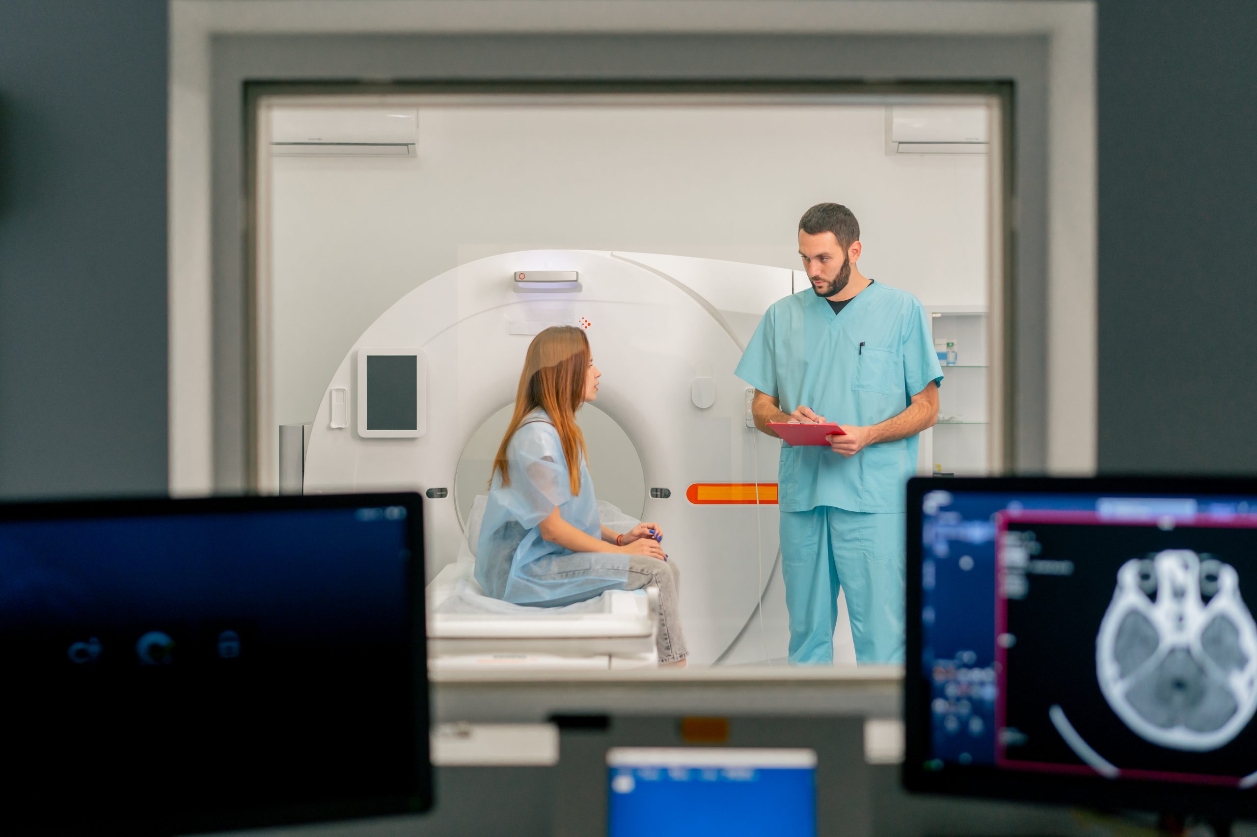
Powered By Innovation At Wayne Radiology Center patients can experience top of the line MR imaging. The 3T High Field  Skyra MRI offers super high definition scanning of musculoskeletal,  neuro, MR angiogram, dynamic prostate, breast MR, abdomen and pelvis MRI with no breath holds, MR enterography, non contrast vascular MRA, diffusion tensor imaging (DTI), arthrograms, cartilage knee mapping. Magnetic Resonance Imaging An MRI is a form of diagnostic medical imaging that utilizes a very large magnet along with radio waves to create cross-sectional images of your internal tissues.  MRI is able to create incredibly accurate images while being totally harmless and non-invasive to the person receiving it. It does not utilize radiation like other forms of imaging, making it 100% safe for use with most people. When it comes to image quality and accuracy, it doesn’t get better than our 3T Skyra Wide Bore MRI at Apex Radiology. How Does an MRI Work? Magnetic resonance imaging (MRI) is a safe and painless test that uses a magnetic field and radio waves to produce detailed pictures of the body’s organs and structures. With our advanced 3T Wide Bore MRI machine you can expect each scan to be completed in less than 15 minutes. WHAT CAN AN MRI IDENTIFY ? MRI can provide highly detailed images of vital organs, muscles, and other soft tissues. Bone does not provide the necessary signal, so they appear as black areas around the soft tissues.  Because of these detailed images, MRI can detect detailed abnormalities in the brain such as stroke, tumors, or infection. TALK TO YOUR DOCTOR BEFORE GETTING AN MRI It can also easily detect issues pertaining to cardiovascular health as well as the nervous system such as spinal problems and various forms of venous disease. MRI should not, however, be used to identify issues with those who are currently using pacemakers, insulin pumps, cochlear implants, or have metal placed somewhere in their body to avoid complications with the device. Book Appointment

ULTRASOUND 2 scaled

Elastography Ultrasound (Fibroscan)

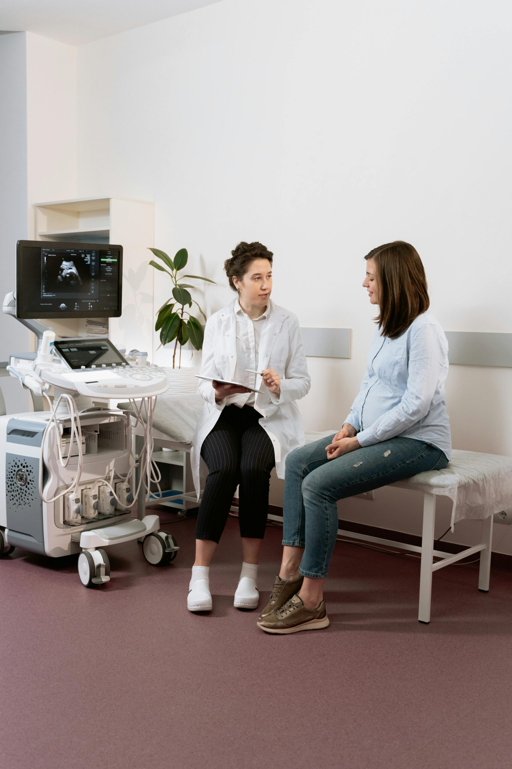
Liver Ultrasound Shear Wave Elastography Experience the future of liver disease diagnosis and monitoring without the need for invasive biopsies. Our advanced medical facility proudly offers liver disease diagnostic services that employ non-invasive techniques, sparing you from the discomfort of traditional liver biopsies. Using state-of-the-art technologies, such as Shear Wave Elastography, we can assess liver health with remarkable precision. This means accurate and detailed insights into the condition of your liver without the need for surgical procedures. Our commitment to innovation ensures that you receive thorough and reliable information about your liver’s well-being, facilitating timely interventions and personalized care plans. Liver Fibrosis and Cirrhosis Cirrhosis of the liver,” which involves loss of liver function due to the formation of scar tissue, may be a term familiar to many. However, what may be less familiar are the many causes of the disease, as well as an earlier, preceding disease stage called fibrosis. In fibrosis, the process of scar tissue formation in the liver has begun but has not yet progressed to the irreversible tissue damage that occurs with cirrhosis. Among the conditions associated with fibrosis and cirrhosisare:● Viral hepatitis● Obesity● Type 2 diabetes● Non-alcoholic fatty liver disease● Excessive alcohol intake The Importance of Early Detection and the Role of Liver Ultrasound Elastography Regardless of cause, detecting liver fibrosis before it has progressed to cirrhosis can make a significant difference in patient outcomes. The good news is that liver ultrasound shear-wave elastography enables radiologists to accurately diagnose liver disease without subjecting the patient to an invasive liver biopsy. Instead of requiring extraction of an actual sample of liver tissue for testing, elastography makes it possible to measure the stiffness of liver tissue through the advanced technology of shear wave ultrasound. Elastography evaluates the degree of stiffness of liver tissue and assigns a score that correlates with the level of fibrotic changes present. Other Benefits of Liver Ultrasound Shear Wave Elastography In addition to the non-invasive nature of the procedure, liver ultrasound shear wave elastography also offers patients the benefits of:● No risk of complications● Ease of repeated exams for patients who need regular monitoring of their liver health● Interpretation by an expert diagnostic radiologist● Prompt reports to the referring physician● Ability to complete the exams at Princeton Radiology’s convenient outpatient imaging centers● Reduced cost Who Is a Candidate?Patients are candidates for liver ultrasound shear wave elastography if they:● Need initial diagnosis or staging of chronic liver disease, such as chronic viral hepatitis or non-alcoholic fatty liver disease● Have known cirrhosis that requires assessment and staging● Have unexplained portal hypertension● Require follow-up after an initial diagnosis of liver fibrosis, to monitor disease progression● Are in treatment for liver disease and need follow-up to monitor response to treatment or identify the need for adjustments to treatment Book Appointment

Magnetic resonance imaging scan or MRI machine device in hospita

3T Wide Bore MRI

Powered By Innovation At Wayne Radiology Center patients can experience top of the line MR imaging. The 3T High Field  Skyra MRI offers super high definition scanning of musculoskeletal,  neuro, MR angiogram, dynamic prostate, breast MR, abdomen and pelvis MRI with no breath holds, MR enterography, non contrast vascular MRA, diffusion tensor imaging (DTI), arthrograms, cartilage knee mapping. Magnetic Resonance Imaging An MRI is a form of diagnostic medical imaging that utilizes a very large magnet along with radio waves to create cross-sectional images of your internal tissues.  MRI is able to create incredibly accurate images while being totally harmless and non-invasive to the person receiving it. It does not utilize radiation like other forms of imaging, making it 100% safe for use with most people. When it comes to image quality and accuracy, it doesn’t get better than our 3T Skyra Wide Bore MRI at Apex Radiology. How Does an MRI Work? Magnetic resonance imaging (MRI) is a safe and painless test that uses a magnetic field and radio waves to produce detailed pictures of the body’s organs and structures. With our advanced 3T Wide Bore MRI machine you can expect each scan to be completed in less than 15 minutes. WHAT CAN AN MRI IDENTIFY ? MRI can provide highly detailed images of vital organs, muscles, and other soft tissues. Bone does not provide the necessary signal, so they appear as black areas around the soft tissues.  Because of these detailed images, MRI can detect detailed abnormalities in the brain such as stroke, tumors, or infection. TALK TO YOUR DOCTOR BEFORE GETTING AN MRI It can also easily detect issues pertaining to cardiovascular health as well as the nervous system such as spinal problems and various forms of venous disease. MRI should not, however, be used to identify issues with those who are currently using pacemakers, insulin pumps, cochlear implants, or have metal placed somewhere in their body to avoid complications with the device. Book Appointment

in an ophthalmology clinic young doctor after an e 2024 11 27 10 50 38 utc scaled

Breast Imaging

Early Cancer Detection Saves Lives It’s estimated by medical experts that 1 in 8 women will develop breast cancer in their lifetime. Though it’s far less likely to ever occur in a man, the odds are still roughly 1 in 883 for it to appear in an otherwise healthy male.  To best combat the appearance of breast cancer, early detection is vital. That’s why Apex Radiology assists individuals with breast imaging services that can help detect, diagnose, and treat cancerous tumors before they have a chance to spread and pose a real threat to your life. Breast Cancer Screening Guidelines The appearance of breast cancer becomes prevalent with age. Mammography and breast imaging techniques also expose women to very small amounts of radiation, similar to X-rays. Therefore, how often a woman should undergo screening depends upon her overall health and age. Different cancer networks and medical experts follow different guidelines for which ages and how often the individual should receive a mammography. The American Cancer Society recommends that all women receive breast imaging starting at age 45, and every year after that until the age of 55. So long as the individual remains in optimal health, you can begin screening every 2 years.  These guidelines are based upon otherwise good health conditions. If you begin to experience any changes in your health or have any other risk factors, your doctor may recommend that you receive breast cancer screenings more frequently. As with any form of medical imaging, your doctor will only use techniques that expose you to safe amounts of radiation, and will only recommend an increase in testing if the images could help protect your health and your life. CAN I JUST GET A CLINICAL BREAST EXAM? A clinical breast exam is performed by your health care provider at the same time of your regular health check-up. They will utilize approved techniques to check for lumps or abnormalities in your breasts and underarm area. The American Cancer Society does not recommend clinical exams as a substitute for breast imaging as it can often lead to false-positive results and provide inaccurate information. BREAST CANCER IN MEN Out of all breast cancer cases, men only make up about 1% per year. However, this still accounts to about 2000 cases a year. If you feel a knot or thickened area in the breast or under the arm, you should see your doctor. Other signs may also include rashes around the nipple, change in breast size, or puckering of the skin. Talk to your doctor if your family has history of breast cancer or enlarged breast tissue among men. Book Appointment