doctor makes ultrasound of pregnancy to patient g 2025 03 15 23 03 06 utc scaled

Echocardiogram

Echocardiogram An echocardiogram is a noninvasive, painless ultrasound exam that allows radiologists and cardiologists at Dignity Health to see the structure of your heart as it pumps. A 3-D echocardiogram provides three dimensional images of your heart. Your cardiologist may ask that you undergo a 3-D echocardiogram to see if your heart has any irregularities in its chambers or if there are problems with the valves, which control the blood flow from one chamber to another. The test may be helpful in making a diagnosis if you have been experiencing signs and symptoms of valve disease, such as:   Light-headedness Chest pain Fatigue Swelling in your feet (edema) Shortness of breath Book Appointment

biracial female doctor using laptop with radiograp 2025 04 03 11 56 30 utc scaled

Bone Density/DEXA

DEXA Scan: Bone Density Test A DEXA scan is an imaging test that measures bone density (strength). DEXA scan results can provide helpful details about your risk for osteoporosis (bone loss) and fractures (bone breaks). Bone Densitometry (DEXA) Osteoporosis is a disease in which bone density decreases, making bones brittle and prone to fractures. It is estimated that at age 50, a woman has nearly a 50% chance of developing an osteoporotic fracture during her remaining lifetime. A woman’s lifetime risk of hip fracture alone is equal to the combined risk of developing breast, uterine, and ovarian cancer—and up to 20% more women who suffer hip fractures die within one year of the fracture than those of a similar age who haven’t suffered a hip fracture. It is therefore important to identify those women who are at risk for developing osteoporosis. Procedure Description DEXA Dual Energy X-ray Absorptiometry (DEXA) is the most accurate and advanced test available for measuring bone density (strength). Its results are reproducible, therefore allowing measurements to be taken over time showing progression of disease or improvement in bone density due to treatment. Minimal radiation (less than 1/20 of a chest x-ray) is used to determine the bone density of the spine, hip or wrist. A DEXA test is more sensitive than ordinary x-rays, more accurate than heel measurements and can diagnose bone loss at an earlier stage. Safe and painless, the 10-minute DEXA scan is the most reliable test to determine even the earliest stages of bone loss associated with osteoporosis. The DEXA machine has a large table with a large arm attached over the top of the table. The patient lies down on the table and the arm scans over the patient’s body while images are taken and sent to a computer screen. Facts More than 10 million Americans have osteoporosis. Four out of five of them are women. Osteoporosis is the cause of over 2 million fractures annually, including vertebral, hip, and wrist fractures. Book Appointment

XRAY MACHINE

X-rays

Effective Diagnostic Radiology X-rays are the most commonly used tool in diagnostic radiology. This is an area of medical imaging meant to assist doctors with locating and identifying underlying illness to guide them to proper treatment. Are X-rays Safe? Many people are hesitant to undergo an X-ray as they do expose your body to a very small dosage of radiation. However, the amount of radiation you experience is small enough that it’s equivalent to what you naturally experience from the natural environment. That being said, medical practitioners are very aware of the risks of too much lifetime radiation and will only recommend X-rays or other radiation-based imaging if the benefits of discovering an underlying problem are overwhelmingly helpful for your health. It would take thousands of X-rays to effectively exceed the limits of what is considered healthy radiation exposure. CONDITIONS IDENTIFIED BY X-RAYS Due to the calcium in your bones, X-rays are able to deliver incredibly accurate images of your bones and any breaks or fractures. However, it can also be used to identify issues with your cardiovascular tissues such as the heart, lungs, and blood vessels. These softer areas effectively appear as shades of grey and can help guide treatment and other various internal procedures. IMAGE-GUIDED TREATMENTS This can be used to help your doctor identify certain conditions and propose treatments that are effective against any identified issues. The advancements in radiology allow doctors to accurately diagnose your condition without performing overly invasive procedures. If your doctor prescribes an X-ray to investigate your health, we’re here to help. Please get in touch to schedule your appointment with us today! Book Appointment

ULTRASOUND

Ultrasound

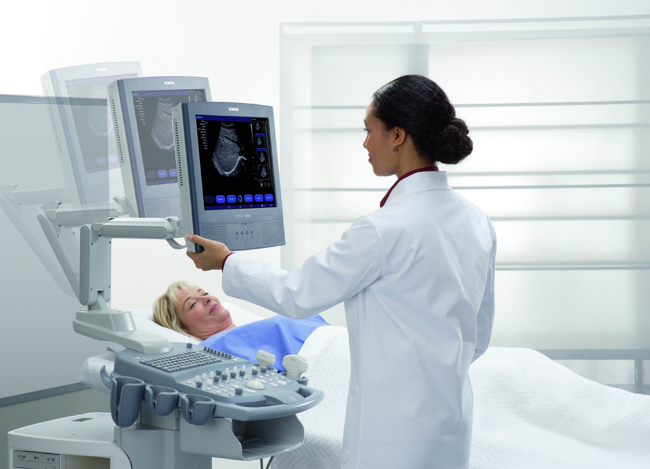
Radiation-Free Medical Imaging Whereas other imaging modalities utilize some dosage of radiation, ultrasounds are conducted utilizing high-frequency sound waves. These sound waves contact internal surfaces and create an image based off of these collisions.   These images can assist your doctor with identifying issues with internal organs and other soft issues without having to open up the body and expose you to traumatic exploratory surgery. What to Expect From an Ultrasound Whenever you are scheduled to receive an ultrasound, you’ll be required to change into a comfortable gown so that the technician can apply the transducer along your skin.  They’ll first apply a lubricating jelly to prevent any uncomfortable friction and allow the transducer to smoothly move across your body and create a clear image. From there, the procedure is painless and requires nothing on your part. The transducer will emit sound waves and create an echo back whenever they strike something inside of your body. Afterward, a medical expert will then carefully examine the images in order to check for any abnormalities or cause for concern. They can then use this information to help prescribe treatment for anything that’s affecting your health. WHAT ULTRASOUNDS DETECT An ultrasound can be used to identify issues with internal tissues, namely those which are mostly solid throughout. The stomach, for example, may not be best seen via an ultrasound due to the open space and air flow. An ultrasound can examine organs such as the gallbladder, liver, or kidneys. It also assists women by identifying issues with pelvic pain. ONGOING PREGNANCY MONITORING Ultrasounds are also used to help monitor your health throughout the duration of pregnancy. It’s a wonderful opportunity to meet your child for the first time, while your doctor monitors the health and growth of the fetus. If your doctor prescribes an ultrasound to monitor your health, we’re here to help. Be sure to schedule your next appointment with us today! Book Appointment