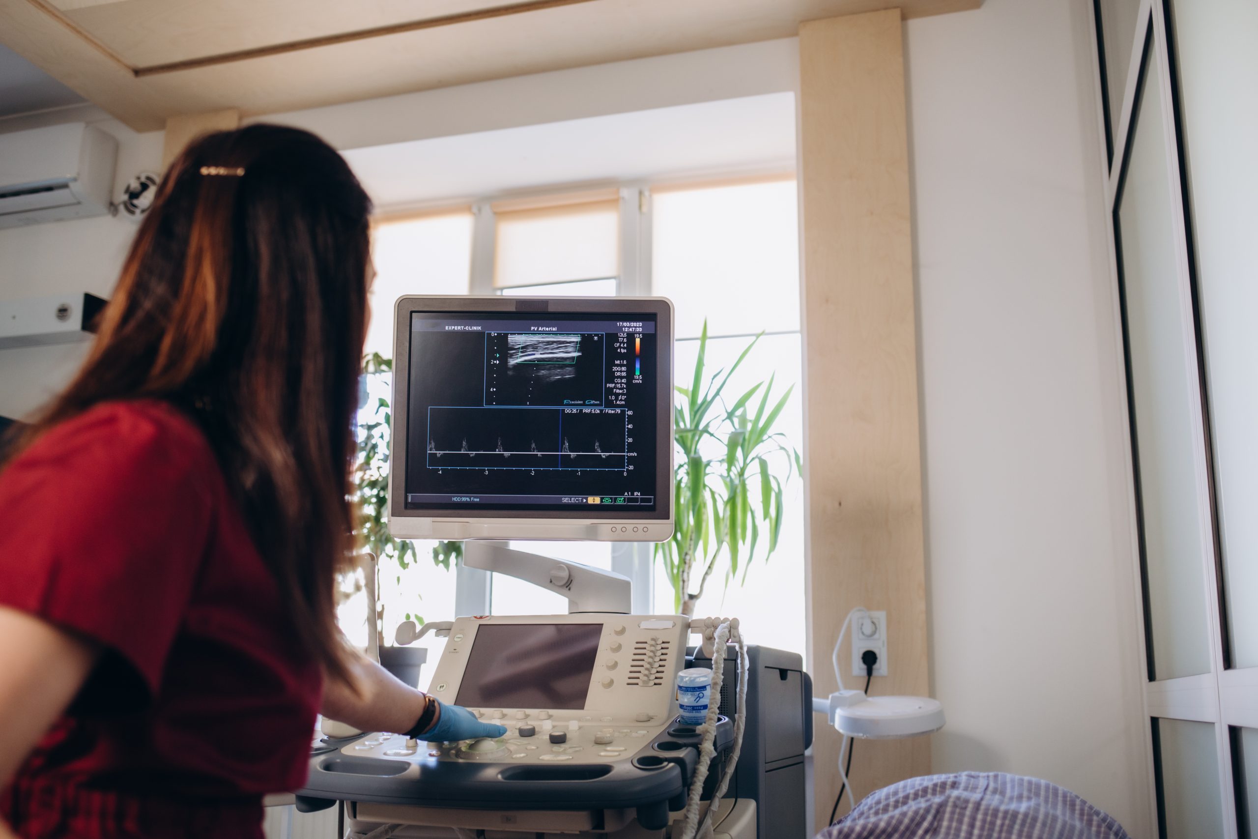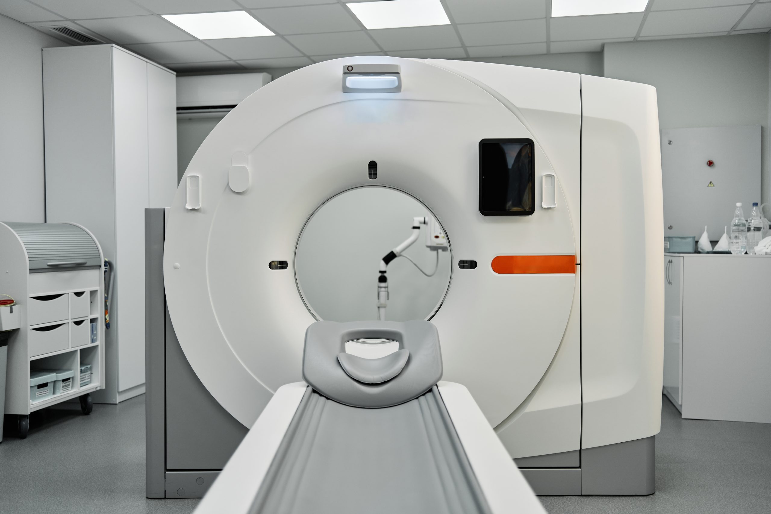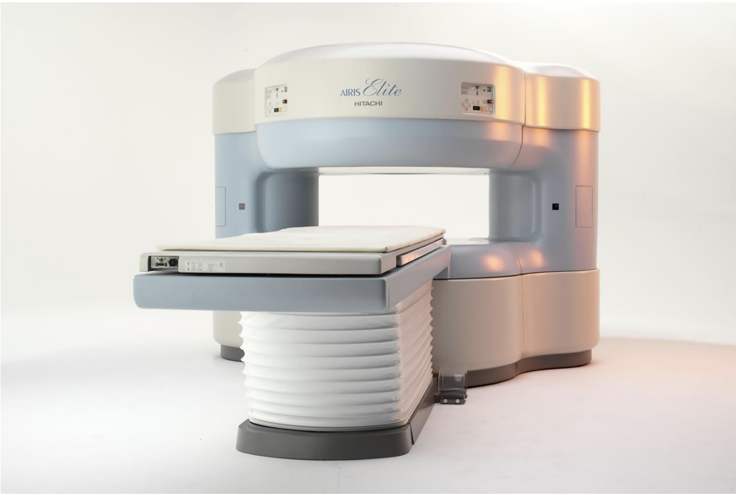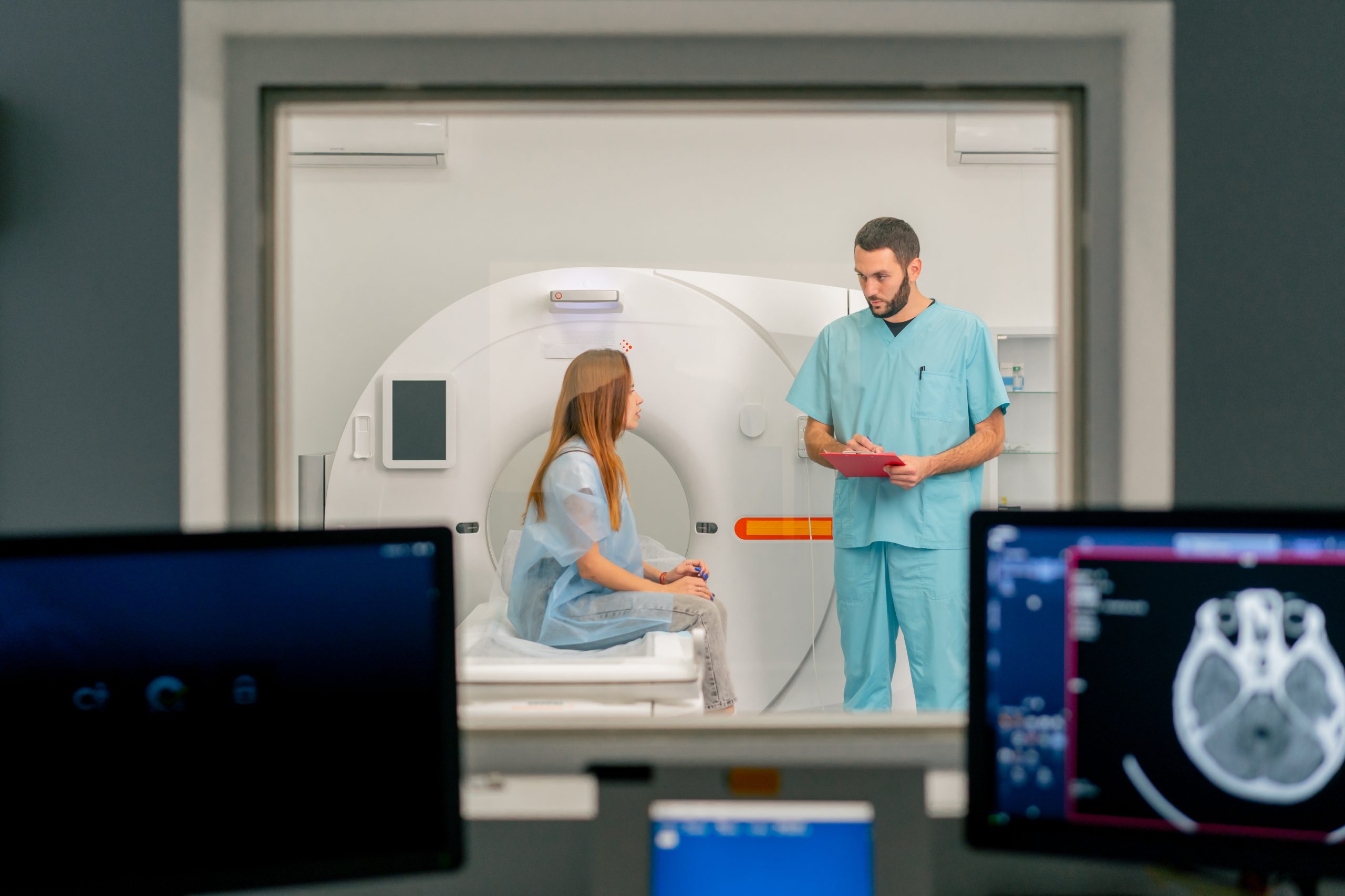
Powered By Innovation At Wayne Radiology Center patients can experience top of the line MR imaging. The 3T High Field Skyra MRI offers super high definition scanning of musculoskeletal, neuro, MR angiogram, dynamic prostate, breast MR, abdomen and pelvis MRI with no breath holds, MR enterography, non contrast vascular MRA, diffusion tensor imaging (DTI), arthrograms, cartilage knee mapping. Magnetic Resonance Imaging An MRI is a form of diagnostic medical imaging that utilizes a very large magnet along with radio waves to create cross-sectional images of your internal tissues. MRI is able to create incredibly accurate images while being totally harmless and non-invasive to the person receiving it. It does not utilize radiation like other forms of imaging, making it 100% safe for use with most people. When it comes to image quality and accuracy, it doesn’t get better than our 3T Skyra Wide Bore MRI at Apex Radiology. How Does an MRI Work? Magnetic resonance imaging (MRI) is a safe and painless test that uses a magnetic field and radio waves to produce detailed pictures of the body’s organs and structures. With our advanced 3T Wide Bore MRI machine you can expect each scan to be completed in less than 15 minutes. WHAT CAN AN MRI IDENTIFY ? MRI can provide highly detailed images of vital organs, muscles, and other soft tissues. Bone does not provide the necessary signal, so they appear as black areas around the soft tissues. Because of these detailed images, MRI can detect detailed abnormalities in the brain such as stroke, tumors, or infection. TALK TO YOUR DOCTOR BEFORE GETTING AN MRI It can also easily detect issues pertaining to cardiovascular health as well as the nervous system such as spinal problems and various forms of venous disease. MRI should not, however, be used to identify issues with those who are currently using pacemakers, insulin pumps, cochlear implants, or have metal placed somewhere in their body to avoid complications with the device. Book Appointment